



TB-500 Research: Full-Length Peptide, Fragment and Evidence

TL;DR · The short version
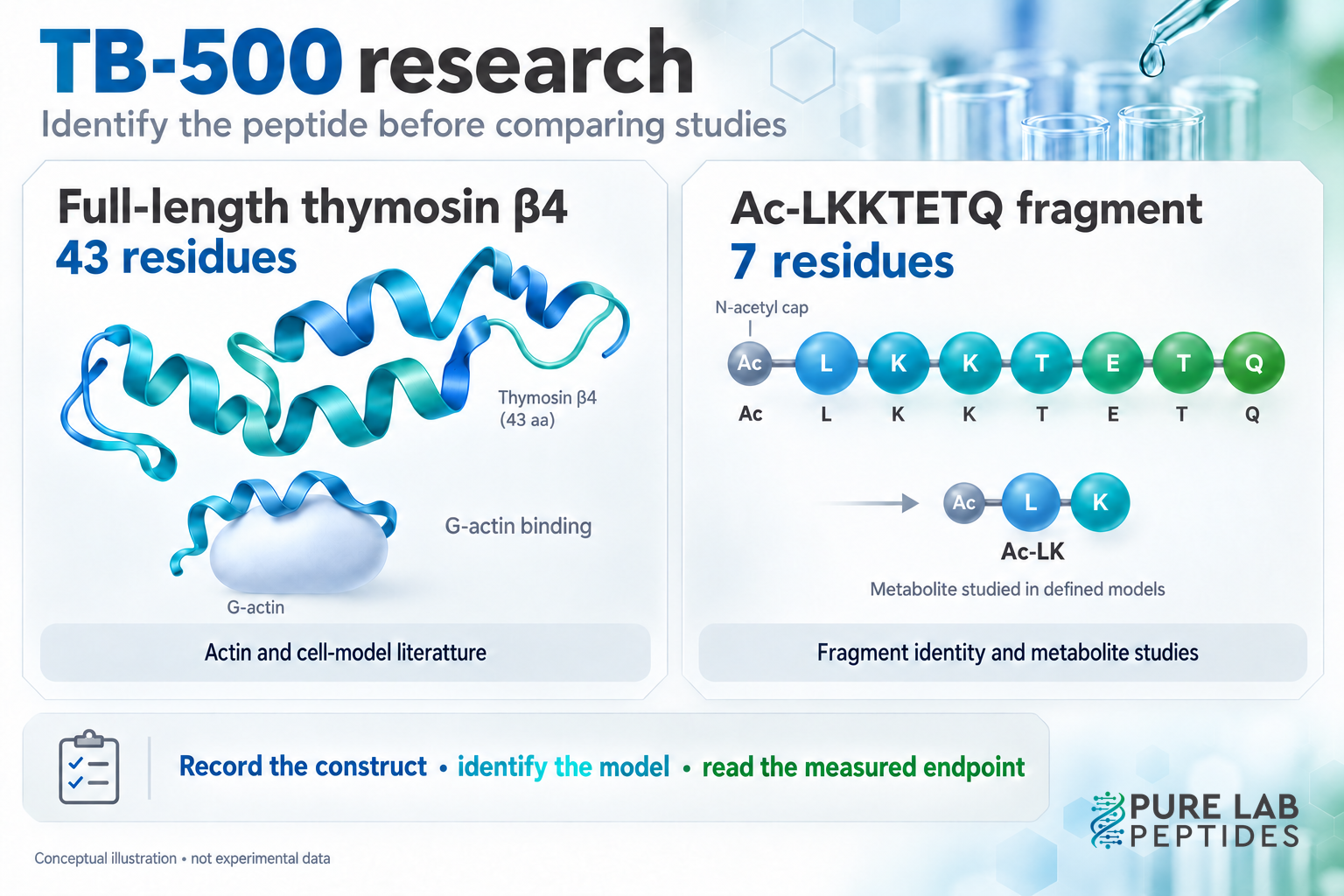
Begin TB-500 research by checking the sequence. Full-length thymosin beta-4 has 43 residues, while several analytical papers call the N-acetylated fragment Ac-LKKTETQ TB-500. The full peptide, fragment and metabolites need separate evidence records.
The name is the snag. Two papers headed “TB-500” may investigate different molecules: full-length thymosin beta-4 in one, seven of its residues in another. A metabolite of the fragment adds another distinction.
Read the material details before the claimed effect. Once you know what was tested, the different strands of this literature are much easier to follow.
Full-length thymosin beta-4 and the short fragment
Quick take: Record the sequence, length and terminal modification. The name TB-500 alone does not resolve every paper’s material identity.
Full-length thymosin beta-4, often written Tβ4, contains 43 amino-acid residues. In 2012, Esposito and colleagues characterized a product called TB-500 and identified its peptide content as the N-terminally acetylated 17–23 fragment of Tβ4: Ac-LKKTETQ. They synthesized the fragment to confirm the analytical assignment. That finding identifies the preparation they examined; it does not characterize every product sold under the same name. Esposito et al., 2012.
Ac marks an N-acetyl cap, not an eighth amino acid. The seven letters LKKTETQ give the residue order. Removing the cap changes the construct even though those letters remain. The sequence notation guide shows how to read that difference.
For our catalogue, the supplier-confirmed TB-500 identity is the full-length 43-residue material associated with CAS 77591-33-4, including the TB-500 component in blends. That catalogue specification is separate from the short-fragment preparations described in the papers below. A registry number does not, by itself, verify the contents of an individual vial.
What full-length thymosin β4 studies measure
Quick take: Actin interaction is one research question. Tissue repair is another. Keep the construct, assay and experimental model attached to each finding.
An original actin study reported that Tβ4 formed a 1:1 complex with monomeric actin under its tested conditions. The complex did not support filament elongation in those experiments. This provides a specific biochemical basis for discussing actin sequestration: binding actin monomers and affecting their availability for assembly. It is not a measurement of clinical recovery. Weber et al., 1992.
Even a small chemical change can matter to interpretation. A 1994 study examined oxidation of methionine at position 6. The oxidized peptide inhibited actin polymerization much less strongly, yet showed similar binding affinity under nonpolymerizing conditions. “Binding” and “polymerization inhibition” were different measurements in that experiment. Thymosin beta-4 oxidation study, 1994.
In a separate rat wound study, Malinda and colleagues examined full-thickness wounds and reported greater re-epithelialization with Tβ4 than saline at the tested time points. The paper also investigated keratinocyte migration in a cell assay. These are preclinical observations with defined models and endpoints. They do not establish recovery from a human tendon injury or the behavior of a seven-residue preparation. Malinda et al., 1999.
How fragment and metabolite research fits in
Quick take: A fragment can have its own analytical and biological literature. Do not quietly transfer those findings to the full-length peptide.
Rahaman and colleagues’ 2024 study defined TB-500 as Ac-LKKTETQ. It developed a mass-spectrometric method for measuring the fragment and metabolites in experimental systems, including rats, and screened wound-healing activity in vitro. The combination of analytical detection and a cell assay is useful, but those parts answer different questions: what was detected, and what activity was observed in that model. Rahaman et al., 2024.
A follow-up published in September 2026 studied the metabolite Ac-LK, N-acetyl-leucyl-lysine. Its experiments included endothelial-cell assays, proteomic profiling and a mouse wound model. Under the tested conditions, Ac-LK showed activity that supported the authors’ hypothesis that metabolic conversion contributes to some effects attributed to the parent fragment. The authors also called for further experiments to establish that mechanism definitively. Rahaman et al., 2026.
These experiments concern the defined fragment and its metabolite. They don’t establish a shared prodrug mechanism for everything named TB-500 or a human effect for full-length Tβ4. The in vitro versus in vivo peptide research helps keep the cell and animal findings distinct.
Measuring TB-500 and its metabolites
The 2024 researchers used UHPLC with high-resolution Orbitrap MS/MS and compared the measured fragment ions with authentic standards. For quantification, they assessed calibration, accuracy, reproducibility, extraction recovery and matrix effects in spiked urine samples. The matrix matters because sample components can change the analytical signal. Original method and validation results.
A labeled peak isn’t enough. Look for the reference standard, matrix and validated range. A concentration method for urine doesn’t automatically answer a dry vial’s purity question.
A comparison table that keeps the molecules separate
Quick take: Give each construct its own row. Then record the model, measurement and comparison before summarizing the result.
| Material examined | Example research | What to keep separate |
|---|---|---|
| Full-length Tβ4, 43 residues | Actin interaction and rat wound experiments | Biochemical binding, cell migration and tissue endpoints |
| Oxidized full-length Tβ4 | Actin polymerization and binding assays | The oxidation state and each assay’s conditions |
| Ac-LKKTETQ, 7 residues | Fragment identification and metabolite measurements | The N-acetyl cap, analytical method and biological model |
| Ac-LK metabolite | Endothelial-cell assays and mouse wound experiments | The tested metabolite, comparison group and limits of the proposed mechanism |
Consider two hypothetical notes: Tβ4 improved wound healing in rats, and Ac-LK changed endothelial-cell behavior. Neither note establishes that a TB-500 vial improves human recovery. Combining them still leaves the molecule, model and outcome to be demonstrated.
Give each construct its own row, then add the model, comparator, time point and endpoint. The research-reading guide expands that checklist, while the mechanisms and consumer claims guide explains why a plausible pathway leaves outcome questions open.
Common TB-500 research questions
Quick take: Resolve identity from explicit material details, not from the paper’s headline or one mass-spectrum peak.
Is TB-500 always the seven-residue fragment?
No universal identity should be assigned from the name alone. The cited analytical papers defined their material as Ac-LKKTETQ; our catalogue uses supplier-confirmed full-length Tβ4. Read the sequence and preparation details for the specific material being discussed.
Can a mass-spectrum peak provide a CAS number?
A peak does not uniquely assign a registry number. Its mass-to-charge ratio also depends on charge state and ion composition. The 2012 characterization used several analytical steps and a synthetic comparison to confirm the fragment, rather than treating one peak as a complete identity certificate.
Can I compare the fragment and full-length peptide as equivalents?
You can compare related constructs, but equivalence is something that comparison must establish. Shared sequence ancestry doesn’t make activity, metabolism or tissue effects identical.