



Short Peptide Bioregulators: Sequences, Studies and Evidence

TL;DR · The short version
Short peptide bioregulators include distinct sequences such as KE, AED, KED and AEDG studied for cellular regulation. Start with the molecule and cell model, then the result. Docking, gene expression and protein markers supply different evidence; the category name doesn’t establish an effect.
The label “bioregulator” is only an introduction. Which sequence was tested, what changed and how was that change measured? Those details make the research useful.
We’ll follow named sequences and original studies rather than treating the group as interchangeable. That makes it easier to distinguish a measured finding from the broader claims attached to short peptides.
What does short peptide bioregulator mean?
The starting point: Treat the term as a research description. Use the sequence to identify the molecule.
In the studies discussed here, the peptides contain two, three or four amino-acid residues. KE means lysine-glutamate; AED means alanine-glutamate-aspartate; KED means lysine-glutamate-aspartate; and AEDG adds glycine to AED. These are distinct sequences, not alternative spellings of the same compound.
A sequence is more informative than an organ-related brand name. A synthetic sequence also isn’t automatically equivalent to a tissue extract described as a peptide complex. An AEDG finding belongs to that preparation and its experimental conditions.
Our sequence notation guide explains how residue letters and chemical modifications help describe a construct. Here, the focus is what the short-peptide studies actually measured.
What can computer docking tell us?
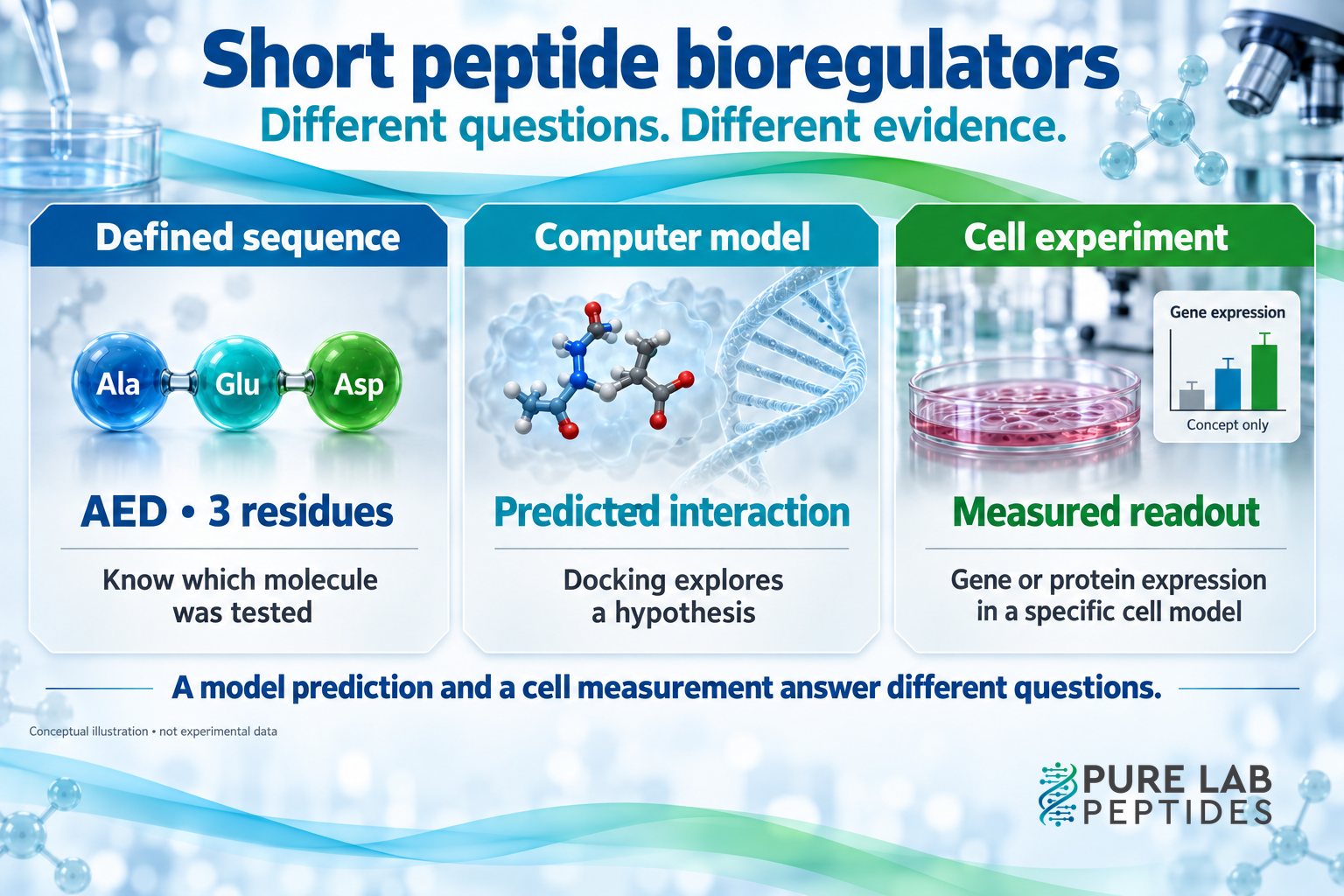
Prediction has a role: Docking can suggest a possible interaction. It does not directly show that the interaction occurred inside a living cell.
The 2016 paper “Short Peptides Regulate Gene Expression” used spatial models of 19 short peptides to explore possible peptide-DNA interactions. The authors proposed that such interactions could help explain effects on gene expression, while noting that the mechanism was unclear.
A title can sound more conclusive than its modeling results. Docking explores a proposed arrangement; cellular entry, direct binding and a target-gene change need appropriate measurements. The image doesn’t establish that whole pathway.
The prediction gives researchers a hypothesis. Keep it described as predicted until experiments support a stronger statement about the interaction.
Why the cell model can change the finding
Context matters: The same peptide and gene can show a different response in different culture-aging models.
Ashapkin and colleagues’ 2020 study tested AED, KED and KE in an embryonic bone-marrow mesenchymal stem-cell line. The researchers used two culture-aging models, described as “passages” and “stationary,” and examined genes associated with the processes under study.
One finding makes the importance of the model clear. KED inhibited TNKS2 expression in the passages model but stimulated it in the stationary model. “KED increases TNKS2” would therefore leave out half of the comparison. “KED decreases TNKS2” would have the same problem.
Retain the gene and culture context when stating the finding. Cell-culture results don’t demonstrate reversal of aging in people, and altered expression doesn’t establish a whole organ’s functional outcome.
Neural markers: what changed, and what remains open?
A marker is a readout: Changes in neural-associated proteins or transcripts are observations in a model, rather than proof of a clinical effect.
A 2019 study of neuronal differentiation investigated AED, KED, KE and AEDG in human periodontal-ligament stem cells. The authors reported higher expression of the markers GAP43 and Nestin with the peptide mixture and with KED alone, using immunofluorescence and Western blotting.
The protein measurements support changes in the reported markers. They don’t independently show fully functional neurons or treatment of a neurological condition.
A separate 2020 AEDG study used human gingival mesenchymal stem cells. It reported changes in neural-associated gene-expression markers and also used docking to explore interactions with histone proteins. The cell measurements and the proposed interaction were different parts of the work. Gingival cells also shouldn’t be silently substituted for the periodontal-ligament cells in the earlier study.
| Original study | Construct or system | Evidence type | Keep this limit attached |
|---|---|---|---|
| Short Peptides Regulate Gene Expression, 2016 | Spatial models of 19 short peptides | Docking predictions involving DNA | Predicted interaction is not observed binding in a living cell |
| Ashapkin and colleagues, 2020 | AED, KED and KE in two culture-aging models | Gene-expression changes | Response depended on the model; no human aging outcome measured |
| Neuronal differentiation study, 2019 | Short peptides in periodontal-ligament stem cells | Protein-marker measurements | Markers alone do not establish mature neuronal function |
| AEDG study, 2020 | Gingival mesenchymal stem cells and separate histone docking | Gene-expression measurements plus modeling | Do not merge a predicted mechanism with a measured cellular result |
How to compare two bioregulator claims without losing the details
Make the comparison specific: Write down the sequence, model, readout and comparison before deciding whether results agree.
Two hypothetical summaries say “supports neural differentiation” and “changes gene expression.” Check their sequences, cells and measurements before calling them corroboration. They may describe different experiments rather than repeated evidence of one effect.
A short note can track peptide, system, measurement and comparator. Specify whether the readout was a transcript, protein marker or functional test, and whether the control was untreated cells, another peptide or another culture condition. That is where the broad description becomes a checkable finding.
Our guide to reading peptide research expands that approach. If the paper combines cultured cells with an animal experiment, the in vitro versus in vivo guide helps keep the models distinct.
Common questions about short peptide bioregulators
A useful boundary: Named short sequences have specific research records. Broad claims about the whole category need their own evidence.
Are AED, KED and AEDG interchangeable?
No. They have different residue compositions or lengths. Match the tested sequence before transferring a result from one article to another.
Do these studies establish that every bioregulator targets one organ?
No. The cited work concerns particular sequences, modeled interactions and cell systems. It does not establish universal organ specificity for every material described as a bioregulator.
Does a proposed DNA or histone interaction prove an outcome in people?
No. A proposed mechanism and a human outcome are separate claims. Our guide to mechanisms and outcome evidence explains that distinction. Start with the actual experiment, then ask what further evidence would be needed for the broader claim.