



Retatrutide Mechanism of Action: Three Receptor Targets

The retatrutide mechanism of action involves one engineered peptide acting as an agonist at three receptors: GLP-1R, GIPR and the glucagon receptor, GCGR. It is also known as LY3437943. The important detail is that three targets do not mean equal activity at each one. Coskun and colleagues’ discovery paper describes that unequal profile.
TL;DR · The short version
Retatrutide isn’t a blend of three hormones. One engineered molecule acts at three receptor targets. When reading an activity result, keep the reference ligand and measured response with the receptor name. A structure can help explain recognition, but it can’t identify the contents of a separately supplied vial.
One molecule, three receptor targets
“Triple agonist” tells you about the targets tested. It doesn’t list three ingredients.
A question like “does it contain GLP-1?” mixes up a molecule with a receptor. Retatrutide can activate the GLP-1 receptor without being a bottle of GLP-1, GIP and glucagon mixed together. The receptor names tell you which targets were tested. They don’t list the ingredients.
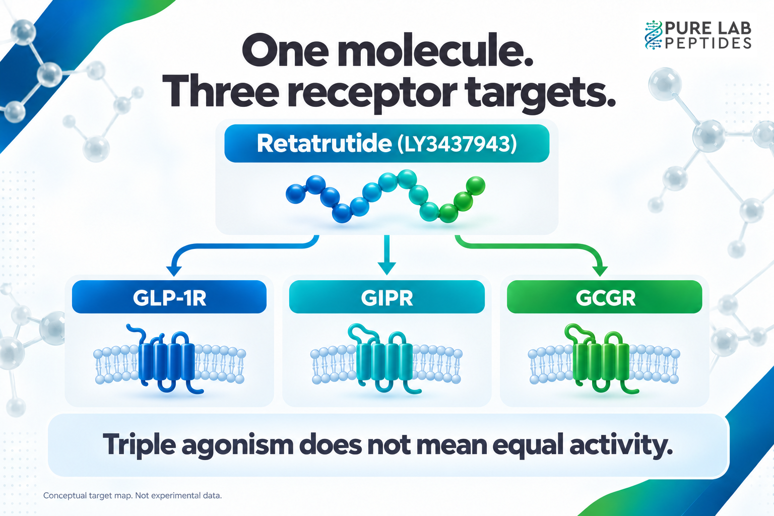
The original discovery work reported activity at all three targets, with greater GIPR activity and a more balanced profile across GLP-1R and GCGR in its in vitro comparisons. That is why “equally strong at three receptors” would be an inaccurate shortcut. The 2022 paper is the starting point for that distinction.
Our GLP-1 receptor agonism guide explains why binding and a functional response aren’t the same finding. With retatrutide, you’ll also need to check how the assays allow comparisons between targets.
Why combine these receptor systems?
The rationale: The GLP-1 receptor, GIP receptor and glucagon receptor connect the molecule to different questions in metabolic research.
GLP-1 and GIP are incretin hormones associated with insulin secretion. In the human comparison by Vilsbøll et al., their insulin responses were measured at controlled glucose levels. Glucagon adds a different question: liver glucose production. Chhibber et al. separated that production into gluconeogenesis and other liver glucose release during a human infusion experiment.
These hormone experiments help explain why researchers would study the receptors together. They didn’t test retatrutide. For its combined profile, use experiments on the engineered peptide itself rather than adding up findings from separate hormones.
Read the receptor result before comparing potency
Start here: Ask “potent at which receptor, compared with what, in which assay?”
Picture a figure with three concentration-response curves. A curve reaching its midpoint sooner looks easy to rank. But first check how it was measured. This is a hypothetical example of reading a figure, not retatrutide data.
Look for the cell type, receptor expression, readout and reference ligand. If each curve was normalized to a different control, equal percentages needn’t represent equal signals. Without those details, the apparent ranking may be answering a question the experiment never asked.
| Field to record | What belongs in it | Why it matters |
|---|---|---|
| Target | GLP-1R, GIPR or GCGR; receptor species and construct | A result belongs to the tested receptor system. |
| Reference | The comparison ligand and normalization rule | “More potent” needs a named comparison. |
| Readout | The measured signal, response timing and baseline | Different endpoints answer different questions. |
| Curve | Concentration range, maximum response and fitted midpoint | A midpoint alone leaves out response size. |
Record the comparison the methods actually support. Counting receptors won’t give you an overall effectiveness score, and “three times the action” leaves out the measurement you need.
What the structural study adds
The extra evidence: Receptor-bound structures help explain how the same peptide can engage different targets.
Li and colleagues’ 2024 study examined retatrutide bound to GLP-1R, GIPR and GCGR using cryo-electron microscopy. The researchers paired those structures with receptor mutations and cAMP measurements. That combination links proposed contacts to a functional test, rather than relying on a molecular picture alone.
Read the structural finding beside the response measurement. Which contact did the researchers propose, and what changed when they tested it? A molecular model alone isn’t an activity curve or an identity test for your sample.
Keep receptor evidence and material evidence separate
Before using the paper: Identify its material, model and endpoint, then identify the evidence for your own sample.
Receptor assays, animal experiments and clinical studies sit at different levels. If a paragraph starts with a cell result and ends with a promise about people, a step has been skipped. Our in vitro versus in vivo guide explains how to keep those levels visible.
For material review, use a separate record: sample identity, analytical method, lot and the document supporting each claim. A purity percentage and a verified molecular identity are different questions, as covered in purity versus identity. Neither one can be filled in from a mechanism diagram.
For your notes, name the compound, receptor, experiment and finding. Keep its comparator and limitation nearby. You’ll have a usable explanation of the mechanism without turning it into evidence for an unmeasured outcome.