



Cell Migration Assays: Scratch, Transwell and 3D Models

TL;DR · The short version
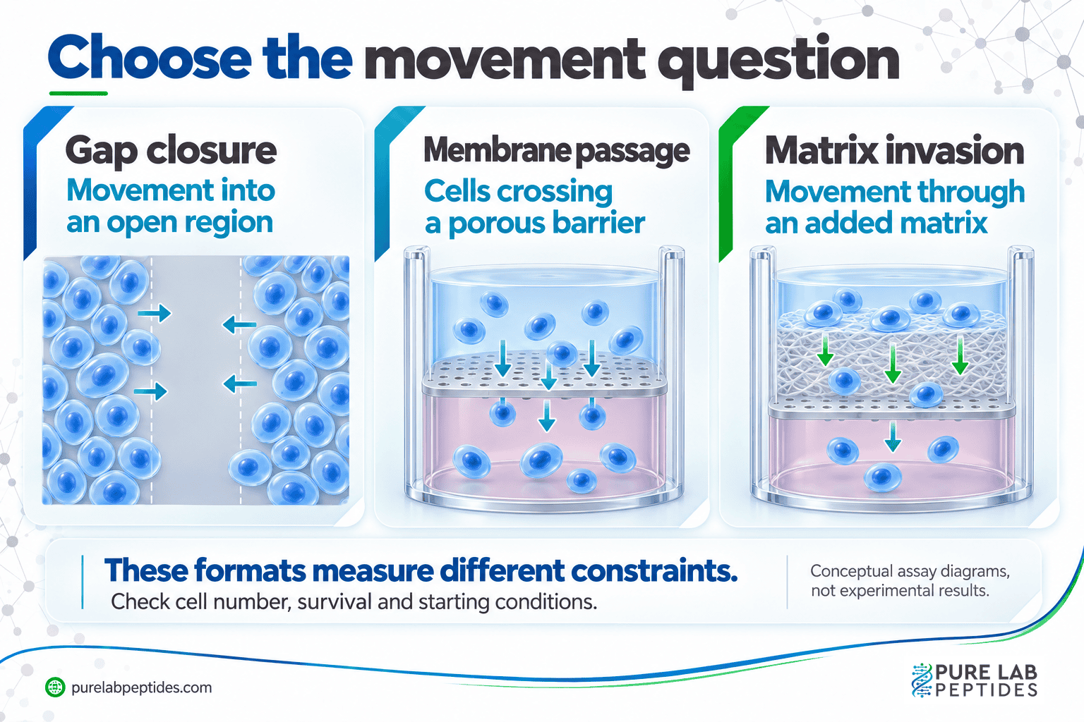
Match the cell migration assay to the movement question. Scratch assays measure net gap closure; transwells measure membrane passage, with matrix barriers adding invasion-related constraints. Starting geometry, division, survival and cell number can affect those readouts too.
The next image shows a smaller gap. Before calling that faster movement, check whether cell division or survival also changed.
The cell migration assay determines what the image can tell you. Scratch, transwell and three-dimensional formats put different constraints on movement. Choose the endpoint first, then interpret it with the cells and controls used.
Match the assay to the movement you want to measure
TL;DR: Choose gap closure, membrane passage, matrix invasion or cell tracking according to the question.
- Gap closure: follow a cell sheet moving into an open region.
- Transwell migration: measure passage through a porous membrane between compartments.
- Matrix invasion: assess movement when a matrix barrier is added.
- Single-cell tracking: examine trajectories when speed and direction matter individually.
A more elaborate format isn’t automatically more useful. It needs to fit the question, and conditions established for one cell line may need adjustment for another.
Scratch or gap-closure assays
TL;DR: Scratch-assay closure can reflect movement, division, survival and starting geometry.
The scratch-assay method described by Liang and colleagues follows closure of a gap in a cell monolayer. Images over time can show changes in collective movement and cell behavior near the edge.
Look at the starting gap as well as the final image. Geometry, attachment, division and survival can contribute to closure. A supported faster-closure result is useful on its own; calling it better healing requires a different evidence base.
Transwell migration and invasion
TL;DR: Report uncoated-membrane migration separately from passage through an added matrix barrier.
Transwell methods examine cells moving through a porous membrane between compartments. A chemoattractant difference can test directional responses. Adding an extracellular-matrix barrier changes the experiment into an invasion-related measurement.
Keep uncoated-membrane migration separate from matrix-coated invasion. To compare counts, also check cell loading, viability, gradient conditions and how the cells were counted.
For example, Justus and colleagues’ published demonstration compared mouse B16F10 melanoma-cell migration toward fibroblast-conditioned medium with migration toward control buffer. The contrast depends on that cell system and stimulus. A count from a single compartment without the relevant comparison would answer less.
What a 3D model adds
TL;DR: A 3D matrix adds physical and biological constraints; it is not automatically a better version of a 2D assay.
A matrix introduces adhesion sites, confinement and a structured route for movement. Those features may suit the physical question, while adding composition and mechanical properties to the comparison.
Before carrying a two-dimensional finding into a 3D model, identify which constraints changed. A similar direction of effect can’t be assumed from the assay names.
Choose a measurement before interpreting the images
TL;DR: Name the readout and its limits before interpreting a representative image.
| Readout | What it describes | What to avoid inferring automatically |
|---|---|---|
| Gap area over time | Net closure of an initially open region | Movement independent of cell division or survival. |
| Cells crossing a membrane | Passage under the assay’s conditions | A specific molecular mechanism. |
| Tracked cell paths | Speed, displacement or directionality | Equivalent behavior in a different matrix or tissue. |
| Movement through a matrix | Behavior under added structural constraints | A clinical invasion or repair outcome. |
Make quantification reproducible
TL;DR: Use a defined calculation and consistent measurement rules; 40% closure is not automatically 40% more migration.
For a gap-area readout, one transparent calculation is percentage closure = (starting area − remaining area) ÷ starting area × 100. As a hypothetical example, an area decreasing from 100 to 60 units is 40% closed. This describes net closure; it does not remove the contribution of cell division.
Keep image scale, region selection and segmentation consistent. Save the starting image and measurement times. For membrane assays, specify cells counted, stained area or calibrated signal; these endpoints don’t share a numerical meaning merely because they track related behavior.
Controls and limitations to check
TL;DR: Check viability, attachment, starting conditions and independent replication alongside movement.
- Compare with the appropriate vehicle or untreated condition.
- Check viable cell numbers and attachment, not only the movement endpoint.
- Keep starting density, gap geometry or membrane conditions comparable.
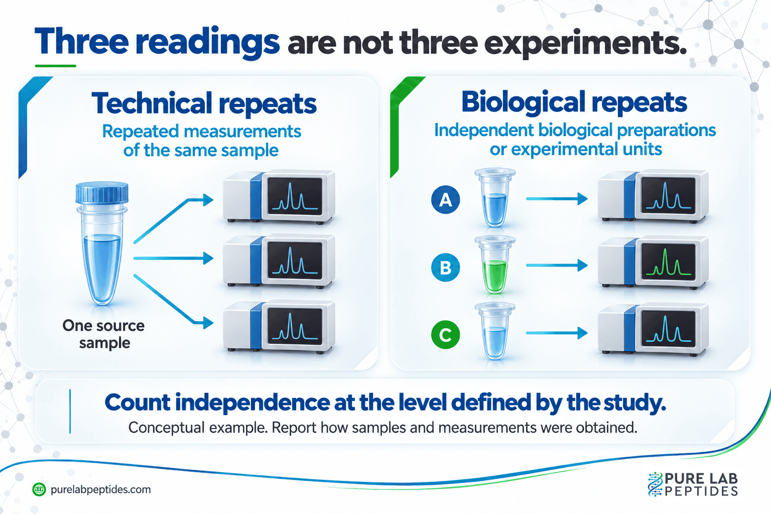
- Report independent biological repeats separately from repeated readings of the same sample.
- Explain any measure used to limit proliferation and whether it changes the cells’ behavior.
The transwell methods paper explains the format’s conditions, not universal settings for every cell line. An endpoint image shows the endpoint. Following the paths cells took requires time-resolved observations.
Interpreting a peptide-associated effect
TL;DR: A peptide-associated change requires its own material and control evidence and does not by itself establish tissue repair.
The published comparison of migration and invasion methods is useful for matching the assay to the question. For a peptide experiment, identify the exact preparation, vehicle and measured endpoint. Include the controls needed to distinguish altered movement from reduced cell number or altered adhesion.
A representative image is one part of the result. Independent biological repeats and consistent analysis make the comparison assessable. If the measurements haven’t separated competing explanations, keep those explanations in the conclusion.
To connect an assay result with its appropriate evidence level, read mechanism of action versus efficacy. A measured change in migration can be a useful finding without establishing tissue repair or a clinical outcome.