



Angiogenesis Assays: Networks, Sprouting and Perfusion Evidence

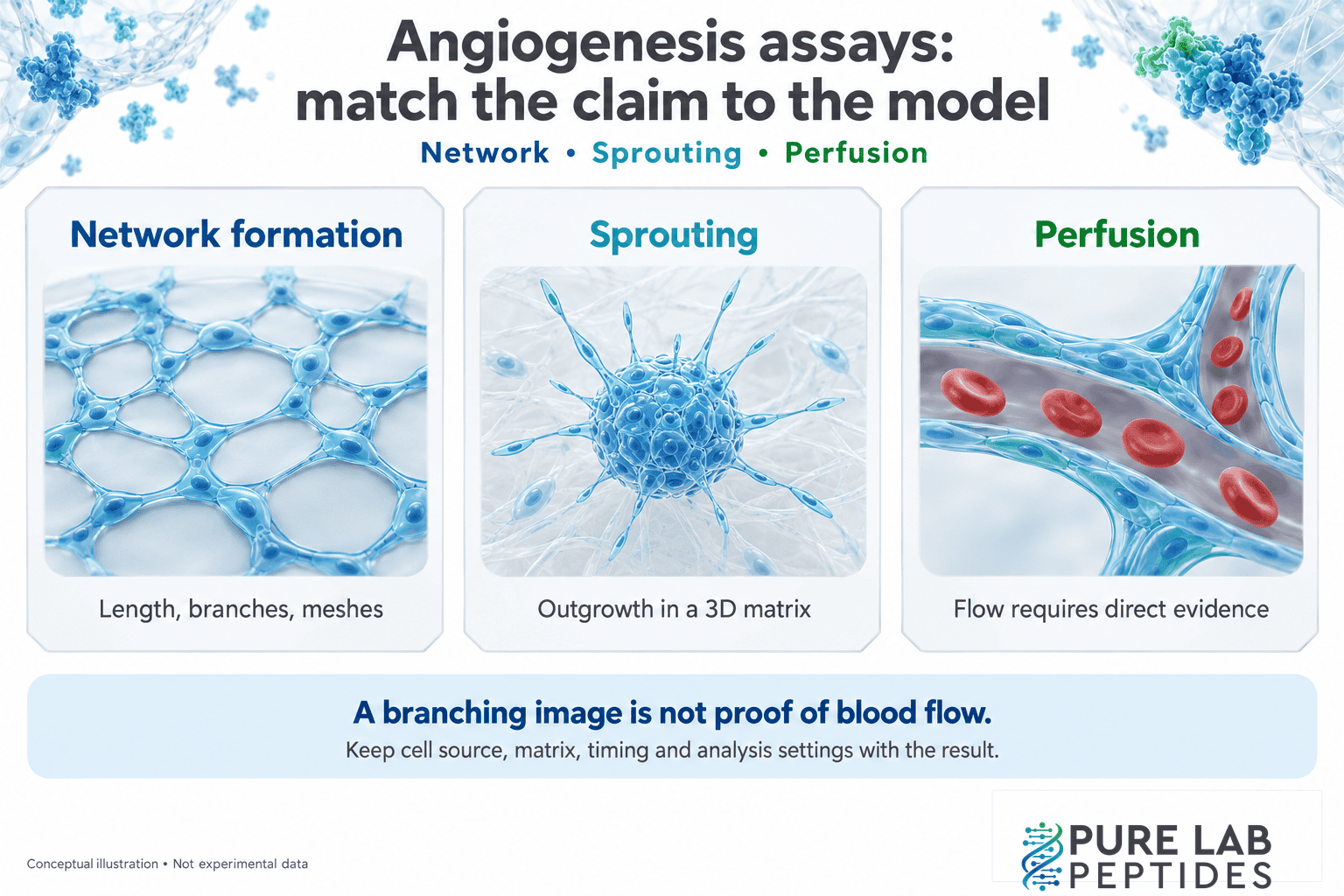
Angiogenesis assays examine how new blood vessels form from existing vessels, using models that capture different parts of the process. Network patterns, matrix sprouting, tissue outgrowth and vascular function are separate measurements. Identify the endpoint before comparing a result.
A branching image shows a pattern. Evidence of blood flow, mature walls or function in an organism needs more than its appearance. Start with the model and ask which of those features was measured.
TL;DR · The short version
- Tube formation assays and sprouting assays provide different views of endothelial behavior.
- Network length, junctions and enclosed mesh area are separate measurements.
- Cell source, culture conditions and extracellular matrix affect the comparison.
- Perfusion requires evidence of flow. It can’t be inferred from branching morphology.
- A peptide-associated change needs suitable controls before it can be assigned a specific mechanism.
Match the angiogenesis assay to the question
Quick take: Choose the biological event first. Then ask whether the model and readout can answer that question.
Movement, proliferation and vascular organization contribute to angiogenesis, but measuring one doesn’t quantify all of them. The consensus guidelines on angiogenesis assays explains how the model and its limitations affect interpretation.
| Model | Useful evidence | Limit to keep visible |
|---|---|---|
| Endothelial tube formation on a matrix | Capillary-like network morphology, length, branches and meshes. | Network appearance alone does not establish blood flow. |
| Spheroid or fibrin bead sprouting | Outgrowth from a defined starting structure into a 3D matrix. | Sprouting and 2D network rearrangement are different endpoints. |
| Aortic ring outgrowth | Vessel-associated outgrowth from a tissue explant. | The model includes several cell types and lacks circulating blood flow. |
| A vascular graft model with perfusion measurements | Evidence that newly formed structures connect with a host circulation. | The result remains specific to the tested animal model and measurements. |
The examples below help with reading those distinctions. They aren’t instructions for running the assays.
Tube formation: what a network image shows
Quick take: An endothelial cell tube formation assay is useful for morphology. Check the matrix and cell context before interpreting the pattern.
HUVECs are human umbilical vein endothelial cells. Under suitable conditions, endothelial cells can organize into capillary-like networks on a matrix. The cell source and setup give a tube-formation image its context.
In Kubota and colleagues’ original study, human umbilical vein and dermal microvascular endothelial cells rapidly formed tubular structures on a basement-membrane-like gel, with little or no proliferation in those conditions. Ultrastructural examination provided additional evidence about lumens and junctions. The gel response differed from cells grown on individual matrix components applied to plastic.
The shape change wasn’t simply a newly divided-cell count. The study also makes the matrix description worth retaining: a basement-membrane-like gel isn’t “just collagen,” and a similar-looking image needn’t carry the same structural evidence.
Xie and colleagues compared HUVEC tube formation on normal and growth-factor-reduced Matrigel while examining a CXCR2 inhibitor. Their analysis used tube length as a readout. Matrix choice changed the experimental comparison. Growth-factor-reduced material is still a particular matrix preparation; the name doesn’t make it interchangeable with every other gel.
Keep cell source, matrix preparation, medium, cell density and observation time in the figure record. When several change between studies, network appearance alone can’t identify which change explains the difference.
Sprouting assays: outgrowth and integrity are different
Quick take: A 3D sprouting result describes outgrowth in its matrix. Separate sprout length from the integrity of the starting structure.
In a spheroid sprouting assay, an aggregate supplies the starting structure and outgrowth enters the matrix. In a fibrin bead assay, cells start associated with beads. These geometries add questions that tube formation assays alone don’t address.
Korff and Augustin’s spheroid study illustrates how specific the conclusions can be. In their collagen-gel model, an RGD peptide affected spheroid integrity and cell detachment, yet did not inhibit primary outgrowth. The comparison included an RAD control. Matrix-dependent mechanical effects also differed between collagen and fibrin.
A summary saying only “changed angiogenesis” would lose the distinction between outgrowth and integrity. Preserve the sequence beside the specific response. The sequence and molecular identity guide explains why related motif names don’t guarantee an identical construct.
Aortic rings and perfusion evidence
Quick take: Tissue outgrowth is valuable evidence, but a growing explant is not a circulating vascular system.
Nicosia and Ottinetti studied microvessel growth from rat aortic rings in fibrin and collagen gels. Growth and regression changed with the matrix and observation period. This model retains tissue context that a single-cell culture does not.
Tissue context adds complexity too. The consensus guidelines notes contributions from other cells and the absence of blood flow in aortic ring assays. Those limits stay relevant when an explant result is summarized.
Perfusion belongs to another evidence category. In Laib and colleagues’ endothelial spheroid graft model, transplanted cells formed networks in immunocompromised mice that connected with the host vasculature and were perfused. The study also examined maturation with mural cells. That provides a specific experimental example of flow and maturation evidence beyond an isolated branching image. It does not establish the same outcome for another material or in people.
Read the image analysis, not just the photograph
Quick take: Name the measured parameter and its units. “More tubes” is too vague to compare across studies.
Carpentier and colleagues compared endothelial tube formation with fibrin bead assays using Angiogenesis Analyzer for ImageJ. Their analysis treated the models as complementary measurements of different biological steps. A method that works for one network geometry isn’t automatically equivalent in the other.
Network length describes extent; junctions describe connectivity; mesh area describes enclosed spaces. These measurements can move differently. Use each name rather than collapsing them into an undefined angiogenesis score.
Check phase contrast or fluorescence microscopy, field selection, scale and image analysis rules. Pixel counts need scale to become physical lengths. Pair the image with its quantification output so another reader can follow the calculation.
Imagine equal total network length in two images, with different junction counts and enclosed spaces. Equal length hasn’t made the architecture identical. Neither measurement, by itself, establishes a functional vessel.
Controls that make the claim easier to assess
Quick take: Check whether the comparison isolates the proposed effect, or leaves cell loss, matrix changes and imaging differences unresolved.
- Baseline: identify the untreated or vehicle comparison and what was held constant.
- Model response: see whether an appropriate reference stimulus or inhibitor established the assay’s response range.
- Cell condition: ask whether viability or cell number changed alongside network morphology.
- Material: record the exact peptide, preparation and identity evidence.
- Analysis: check replicate structure, field-selection rules and which parameter supports the conclusion.
A growth factor such as VEGF gives the study a defined stimulus or comparison. Its presence doesn’t make unlike cultures equivalent. Inspect the actual controls rather than assuming the paper performed every check listed here.
If the central question is how cells migrate, the cell migration models guide covers that narrower topic. Analytical purity is a separate question again: batch-specific COA results describe material testing, not its angiogenic activity.
Angiogenesis assay questions
Quick take: Keep the conclusion tied to the model, endpoint and supporting measurements.
Are more branches always a better result?
“Better” depends on the research question. Branching, survival, sprouting and perfusion are different endpoints. Report the measured change before assigning a broader value judgment.
Does a Matrigel network prove that blood vessels formed?
It provides evidence about network formation under the tested conditions. Claims about lumens, maturation or flow need their own supporting measurements.
Can one in vitro angiogenesis assay establish a peptide’s mechanism?
It can show a response in that model. Assigning a mechanism requires additional evidence that separates the proposed molecular action from alternative explanations.