



AMPK Pathway: Energy Signals, Substrates and Study Results

The AMPK pathway links cellular energy sensing with metabolism, growth and autophagy. Its central enzyme, AMP-activated protein kinase, is a three-part complex. Follow a paper from the upstream change to the kinase measurement and then to the downstream response; those are separate pieces of evidence.
A stronger phospho-AMPK band tells you something about that signal. It doesn’t measure the whole energy state, lipid metabolism or autophagy. Keep that limit in view when following a pathway diagram.
TL;DR · The short version
- AMPK has α, β and γ subunits. Nucleotide binding and upstream kinases help regulate its activity.
- AMP and ADP do not produce identical effects on the complex.
- Keep AMPK phosphorylation, substrate phosphorylation and functional measurements separate.
- A response to AICAR alone does not establish that AMPK caused it.
What the AMPK signaling pathway measures
Quick take: Separate the energy input, the kinase state and the measured output before interpreting a pathway claim.
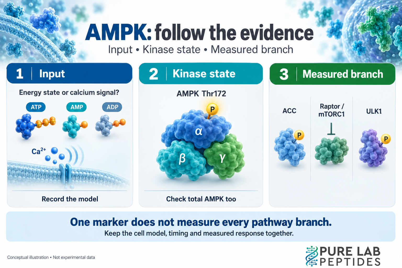
“Energy sensor” is a useful introduction to AMPK, but it can obscure what a study measured. Nucleotide-dependent enzyme activity and acetyl-CoA carboxylase phosphorylation concern different parts of the pathway. The methods tell you which part the result describes.
The AMPK complex is heterotrimeric: α is the catalytic subunit, while β and γ are regulatory subunits. The γ subunit contains nucleotide-binding sites involved in sensing AMP, ADP and ATP. Xiao and colleagues’ structural and biochemical study explains how that binding helps regulate the complex.
For a first reading, label the evidence in the margin:
- Input: an energy-related or calcium-related signal.
- Kinase: AMPK phosphorylation or a direct activity measurement.
- Substrate: a measured protein downstream of AMPK.
- Function: the cellular process the authors actually tested.
These labels are a reading aid. They don’t imply that every paper measured all four.
AMP, ADP and ATP: similar names, different regulation
Quick take: AMP can support allosteric activation; ADP protection against dephosphorylation is a different mechanism.
Allosteric activation means changing enzyme activity through binding at a regulatory site. Phosphorylation adds a phosphate group to a protein. Dephosphorylation removes it. A study can distinguish these events even when a simplified diagram draws one arrow.
In the Xiao study, AMP promoted AMPK phosphorylation, protected against dephosphorylation and produced allosteric activation. ADP also protected against dephosphorylation but did not allosterically activate the complex. Competition with ATP contributed to the nucleotide-dependent regulation.
“AMP or ADP activates AMPK” leaves out the mechanism. Check the nucleotide and preparation, then distinguish the measured events. A phospho-protein result alone doesn’t directly measure ATP production or consumption, even if the authors propose an energy-related explanation.
Upstream kinases: LKB1 and CaMKK2
Quick take: Activation of AMPK can involve different upstream inputs. Identify the route tested in the paper.
Phosphorylation at threonine 172, usually written Thr172, occurs within the activation loop of the α subunit. Woods and colleagues identified LKB1, or liver kinase B1, as a major upstream AMPK kinase. Their work combined a rat-liver preparation, enzyme experiments and cellular tests. This established an important part of the kinase cascade.
But LKB1 isn’t the only relevant input. A later Woods study identified calcium/calmodulin-dependent protein kinase kinase β, now commonly called CaMKK2, as another upstream kinase. Overexpression, inhibition and RNA interference supported its role in the studied systems.
In a calcium-related experiment, increased AMPK phosphorylation needn’t mean the cell ran short of energy. That upstream explanation needs evidence of its own.
AMPK substrates: ACC, mTORC1 and ULK1
Quick take: A downstream branch needs a branch-specific measurement. One marker cannot stand in for the whole pathway.
The role of AMPK in energy homeostasis involves several branches. Here are three useful examples, each supported by a different original study.
| Branch | What the study established | What to check in another paper |
|---|---|---|
| ACC and lipid metabolism | Fullerton et al. examined mice with phosphorylation-resistant ACC1 and ACC2. The mutations altered lipid synthesis and fatty acid oxidation. | Which ACC isoform and phosphorylation site were measured? Is there a functional lipid measurement too? |
| Raptor and mTORC1 | Gwinn et al. showed direct AMPK phosphorylation of raptor and its contribution to mTORC1 inhibition during energy stress. | Did the experiment test the raptor connection and mTORC1 activity, or only AMPK? |
| ULK1 and autophagy | Egan et al. linked AMPK-dependent ULK1 regulation with mitochondrial homeostasis through biochemical and genetic experiments. | Does the study measure a phosphorylation event, autophagy-related markers or a functional process over time? |
The ACC example is especially concrete. The phosphorylation sites tested were mouse ACC1 Ser79 and ACC2 Ser212. Mice carrying alanine substitutions at both sites had increased lipid synthesis and reduced fatty acid oxidation. That is stronger mechanistic evidence than finding a phospho-AMPK band beside a lipid result. Keep the species and site numbering attached to the finding.
Likewise, the raptor study tested specific sites, Ser722 and Ser792, and their role in 14-3-3 binding and mTORC1 inhibition. A summary saying “AMPK regulates growth” leaves out the tested connection. For ULK1, the genetic and starvation experiments provided evidence beyond a single phosphorylation measurement. They do not make every AMPK marker a measurement of autophagic flux.
Why an AICAR response is not enough
Quick take: A reagent-associated effect and an AMPK-dependent effect are different conclusions.
AICAR is frequently used in AMPK research. The ribonucleoside can be converted inside cells to ZMP, an AMP mimetic. It is a small molecule, not a peptide.
That association doesn’t settle every mechanism. Philippe and colleagues studied AICAR in mouse embryonic cells lacking both AMPK α1 and α2. They still observed effects involving LATS1/2 and the Hippo pathway. Their result provides a clear example of an AMPK-independent response in that model.
Look for controls that distinguish a reagent-associated effect from an AMPK-dependent one. Genetic loss of function, rescue and direct activity measurements can address different links. Calling the reagent an activator doesn’t make every subsequent change depend on AMPK.
A practical record for comparing AMPK studies
Quick take: Write down the model, the input and the measured branch. Compare conclusions only after those details match.
Use this short record when two papers seem to disagree:
- Model: species, cell or tissue source, and the reported AMPK isoform or subunit.
- Context: nutrient conditions, stimulus and observation time.
- Kinase evidence: Thr172 phosphorylation, total AMPK and any direct activity measurement.
- Branch evidence: the substrate, site and functional readout.
- Causal check: the comparison supporting AMPK dependence.
Consider two hypothetical reports: one measures phospho-AMPK and another phosphorylated ACC. They might fit the same proposed mechanism, but they aren’t duplicate observations. Neither establishes fatty acid oxidation unless that outcome was measured.
Keep material identity in another record. A pathway result doesn’t verify a supplied vial. Our peptide sequence and identity guide covers molecular descriptions, and the COA results library links analytical documentation to batches.
AMPK pathway questions
Quick take: Use the most specific conclusion the measurements support.
Does more phospho-AMPK mean the whole pathway is more active?
It supports a finding about the phosphorylation signal in that sample. Total protein, activity measurements and downstream evidence determine how far the conclusion can extend.
Are AMPK activation and autophagy the same result?
No. They refer to different events. The ULK1 study provides a mechanistic connection, but a new experiment still needs evidence for its claimed autophagy response.
Can an AMPK paper establish a benefit in people?
A purified-enzyme, cell-culture or mouse finding describes its tested system. It does not establish a human outcome or certify a research material for human use.